Understanding Fluorophores: Excitation, Emission & Fluorescence Spectra
SciFluor explores fluorophores, excitation and emission, fluorescence spectra, brightness, photostability, and their role in modern biological imaging.
Understanding Fluorophores: Excitation, Emission & Fluorescence Spectra
Fluorophores are the molecular components that transform photonic interactions into measurable biological signals. Understanding how they absorb light, emit fluorescence, and respond to their environment is essential for selecting labels and interpreting modern fluorescence images.
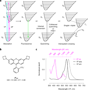
Figure 1 : Fluorescence basics and molecular photophysical processes. Click the figure to view the original Nature Methods article.
Why Fluorophores Matter in Biology
Many biological structures cannot be observed directly with conventional light microscopy because they are too small, too transparent, or lack sufficient optical contrast. Fluorophores provide a controllable optical signal that can be associated with specific biological targets.
A fluorescent molecule may be attached to a protein, nucleic acid, membrane, organelle, metabolite, antibody, or other molecular target. Fluorescent proteins can also be genetically encoded inside living cells, creating a direct connection between molecular biology and optical imaging.
However, fluorescence is much more than a visible color. It is a photophysical process involving absorption, electronic excitation, relaxation and emission. These molecular events determine how a fluorophore behaves inside a microscope and how much information can ultimately be extracted from the resulting image.
1. What Is a Fluorophore?
A fluorophore is a molecule or molecular group capable of absorbing light and subsequently releasing part of that energy as fluorescence. Fluorophores can be naturally occurring, genetically encoded, chemically synthesized, or incorporated into larger fluorescent probes.
In biological research, fluorophores are particularly useful because they can be connected to targeting molecules. A dye may be attached to an antibody, peptide, nucleic acid, ligand or protein tag. This creates an optical signal associated with a particular biological target.
Organic Dyes
Small synthetic molecules with tunable spectral and chemical properties.
Fluorescent Proteins
Genetically encoded reporters used to visualize proteins and cellular processes.
Fluorescent Probes
Molecular systems designed to produce fluorescence in response to a specific target or environment.
2. Excitation: Giving a Fluorophore Energy
Fluorescence begins when a fluorophore absorbs a photon with an appropriate energy. The absorbed energy promotes the molecule from a lower electronic state to an excited state.
Because molecular structures have specific electronic configurations, different wavelengths are absorbed with different efficiencies. This creates characteristic excitation behavior that can be measured spectroscopically.
In microscopy, the choice of excitation wavelength is therefore important. Illumination that efficiently excites a fluorophore can produce a stronger signal while reducing unnecessary exposure of the sample to light.
The wavelength of incident light that provides energy to the fluorophore and initiates its excited state.
3. Emission: Turning Energy Into a Signal
After excitation, a fluorophore undergoes molecular relaxation before returning toward its ground state. Some energy is lost through non-radiative processes, and the remaining energy can be released as fluorescence.
The emitted photon generally has lower energy and a longer wavelength than the excitation photon. This difference is one of the fundamental properties that makes fluorescence microscopy possible.
Optical filters and dichroic elements can exploit the difference between excitation and emission wavelengths to separate the fluorescent signal from the illumination light.
Excitation
Incoming light supplies energy to the fluorophore.
Emission
The fluorophore releases part of its energy as emitted light.

Figure 2 : Fluorescent labeling strategies and examples of small-molecule fluorescent labels.
4. Understanding a Fluorescence Spectrum
A fluorescence spectrum provides much more information than the apparent color of a fluorophore. It describes how excitation or emission changes across wavelength and allows researchers to compare fluorescent labels quantitatively.
Spectral information becomes especially important in multicolor imaging. If the emission spectra of two fluorophores overlap strongly, fluorescence detected in one channel may contain signal originating from another label. This phenomenon can complicate image interpretation.
| Property | What it tells us |
|---|---|
| Excitation maximum | Where excitation is especially efficient. |
| Emission maximum | Where emitted fluorescence is strongest. |
| Spectral width | How broadly the fluorophore responds across wavelength. |
| Spectral overlap | Potential cross-talk between fluorescence channels. |

Figure 3: Examples of fluorophores distributed across the visible spectrum, illustrating the diversity of fluorescent labels.
5. Brightness Is More Than Color
A fluorophore that appears strongly colored is not necessarily the best choice for microscopy. Fluorescence brightness depends on several molecular and experimental properties.
Two important concepts are the extinction coefficient and quantum yield. The extinction coefficient describes how strongly a fluorophore absorbs light, while quantum yield describes the fraction of absorbed photons that result in fluorescence emission.
A useful conceptual relationship
Molecular brightness ≈ extinction coefficient × quantum yield
In real imaging experiments, observed brightness is also influenced by illumination intensity, labeling efficiency, optical collection, detector sensitivity, background and acquisition settings.
6. Photostability: How Long Can Fluorescence Last?
Fluorescence is not necessarily permanent. Repeated illumination can cause fluorophores to undergo photochemical changes that reduce or eliminate their ability to fluoresce. This process is commonly described as photobleaching.
Photobleaching can become an important limitation during long time-lapse experiments, high-intensity imaging and single-molecule measurements. Therefore, selecting a fluorophore involves considering not only its spectral properties but also how stable its signal remains under the conditions of the experiment.
Brightness
Important for detecting weak biological structures and signals.
Photostability
Important when fluorescence must be observed repeatedly over time.
Low Background
Improves the ability to distinguish biological signal from unwanted fluorescence.

Figure 4 : Chemical structures and important considerations associated with small-molecule fluorescent dyes.
7. Choosing Between Fluorescent Proteins and Chemical Dyes
Fluorescent proteins and synthetic dyes provide different strategies for visualizing biology. Fluorescent proteins can be genetically encoded, making them powerful tools for following specific proteins or cellular populations over time.
Chemical dyes can provide high brightness, diverse spectral properties, and specialized chemical behavior. They can also be connected to antibodies, ligands, nucleic acids, or engineered labeling systems.
The choice therefore depends on the biological question, the imaging platform, the required spatial and temporal resolution, and the desired experimental duration.
| Feature | Fluorescent proteins | Chemical dyes |
|---|---|---|
| Labeling | Genetically encoded | Chemical or affinity-based |
| Brightness | Varies between proteins | Often highly tunable |
| Typical applications | Live-cell genetic reporters | Targeted molecular labeling |
8. Fluorophores in Multicolor Imaging
Modern microscopy often attempts to observe several biological targets within the same specimen. Each target can be assigned a different fluorophore, creating multiple imaging channels that can later be combined.
However, adding more colors also increases experimental complexity. Excitation and emission spectra can overlap, and the detected signal may contain contributions from neighboring channels.
Careful fluorophore selection, optical filtering, appropriate illumination and controlled acquisition settings are therefore essential for reliable multicolor imaging.
More colors do not automatically mean more information
The value of multicolor imaging depends on whether individual fluorescence signals can be separated, quantified and correctly assigned to their biological targets.

Figure 5 : Photobleaching and photochemical changes affecting fluorescent dyes during illumination.
9. From Fluorophore Properties to Biological Measurements
The fluorophore is only the beginning of the imaging chain. Once a fluorescent label has been selected, the biological experiment determines how the resulting signal should be interpreted.
Researchers may measure fluorescence intensity, localization, distribution, dynamics, colocalization or changes over time. These measurements can provide information about molecular organization, cellular behavior and biological processes.
However, an increase in fluorescence intensity does not automatically mean that more molecules are present. Fluorescence can also change because of the local environment, illumination conditions, labeling efficiency, photobleaching or detector settings.
10. The Future of Fluorophores
Fluorophore development is increasingly driven by biological requirements. Researchers are designing labels with improved brightness, greater photostability, controlled activation, specific molecular targeting, environmental sensitivity and spectral properties suited to advanced microscopy.
These developments are closely connected to new imaging technologies. Super-resolution microscopy, single-molecule imaging, live-cell imaging, multiphoton microscopy and advanced fluorescent biosensors all place different requirements on fluorescent labels.
Rather than searching for one universally superior fluorophore, modern fluorescence research increasingly focuses on matching molecular properties to the biological question and the imaging platform.
From Molecular Photophysics to Biological Insight
A fluorophore is not simply a colored molecule. Its excitation, emission, brightness, stability, chemical environment and interaction with the microscope all influence the information that ultimately reaches the detector. Understanding these connections allows fluorescence to move from a visual technique toward a quantitative scientific measurement.
Scientific Reading
The following reference provides a detailed scientific overview of small-molecule fluorescent labels, their chemistry, photophysical properties and practical considerations in fluorescence microscopy.
Fluorescence Begins With the Molecule
Explore how fluorophores, spectra, microscopy and quantitative imaging connect molecular behavior with biological discovery.
Explore More Scientific Insights